Reproduction of cells:
Production of gametes for sexual reproduction
Meiosis (My-oh'-sis) is the process by which a single diploid cell splits into four haploid cells called gametes in preparation for sexual reproduction of an organism.
Recall that a diploid cell contains two nearly-identical copies of each chromosome, one from each parent, called a homologous pair of chromosomes.
Human cells, for example, contain 23 different kinds (sorted by length) of chromosomes, and a normal diploid human cell contains two copies of each chromosome, a homologous pair, one inherited from the mother and the other from the father.
So a normal diploid human cell contains 46 chromosomes in all.
Meiosis is a two-phase, multi-step process by which gametes, which will eventually combine to form the first cell of a new organism, are formed.
Gametes are haploid cells. Human gametes contain 23 chromosomes, one of each. Two gametes (one from father, one from mother) will eventually fuse together to form the first diploid cell of the new organism, which will then proceed to divide by mitosis.
In this section we'll go through the steps of meiosis one-by-one, then put them in perspective.
First: chromosome terminology
Different organisms deploy chromosomes in slightly different ways, both in cells and during replication of cells. Here we'll focus on diploid cells (humans have diploid cells), which contain two slightly different copies of each chromosome, forming what is called a homologous pair. Humans have 23 kinds of chromosomes (mostly categorized by their length, from longest to shortest), and there are 2 versions of each kind, making 23 homologous pairs or 46 total chromosomes.
In this section and the section on mitosis, we'll employ schematic drawings of chromosomes that look like this:

On the left is a homologous pair. One came from each parent, thus the ♂ and ♀ symbols. These drawings represent chromosomes that are maximally condensed. The DNA that forms a chromosome is capable of being loosely-coiled — the configuration optimal for use when the cell is performing its principal functions — or tightly coiled and compacted in preparation for cell replication. The coiled form ensures that extremely long DNA molecules can be efficiently segregated into "daughter" cells without tangling.
Here is an electron micrograph of an actual human chromosome during prophase of mitosis:

The pinched-off region is called the centromere. It is the point at which microtubules in the cell attach in order to align and segregate chromosomes into daughter cells.
Before cells can begin mitosis or the first stages of meiosis, protein machinery in the nucleus must make a copy of each chromosome, forming a tetraploid cell, a temporary state necessary for cell division to begin (right side of the panel above).
Meiosis I – Formation of genetically distinct diploid cells
Meiosis begins in the S phase and G2 phases of the cell cycle. In those phases the DNA is replicated in preparation for division either by mitosis or meiosis. The body at the top of this cell is a centrosome, a crucial structure from which microtubules will reach out and attach to the centromeres of chromosomes in order to align and segregate them.
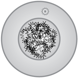
After DNA replication, the cells of an organism that are normally diploid — that occur in homologous pairs — now contain two copies of each chromosome, or two homologous pairs. Before meiosis begins the DNA is in an extended form. It will begin to contract into compact chromosomes, visible in a light microscope, as cell division begins.
Prophase I
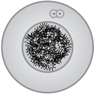
Meiosis is divided into two stages that we label I and II. In prophase I the chromosomes condense into their most compact form. They pair up in groups of four, two pairs of homologous chromosomes, shown here as gray and a magenta pairs of the same length. This hypothetical cell only has three types of chromosomes, just to make the process easier to understand.

The paired homologous pair copies cluster closely as the nuclear membrane dissolves, and the centromeres have replicated and begin to form the microtubules that will eventual surround chromosomes and attach to centromeres. These are the chromosome traffic-control structures.
Finally, a very important process called crossing over occurs between nearby homologs. Segments of DNA are excised and swapped between chromosomes by a set of specialized enzymes. This process effectively mixes up the DNA of both parents of the cell, creating new genetic diversity in the species.
Crossing over is one of the most important sources of genetic diversity in organisms. Here the crossing over is shown by swapping of colors between homologous pairs. In reality many more sections may be swapped. Crossing over is a mixing of the genetic material inherited by each parent from its parents.

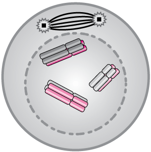
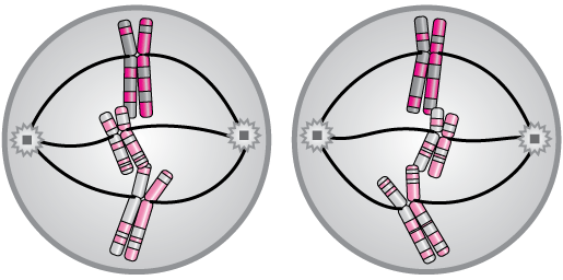
Metaphase I
Once crossing over is complete, the pairs of homologous pairs line up along the metaphase plate, an imaginary plane across the center of the cell. The nuclear membrane has, by now, dissolved. Spindle fibers extend from the centrosomes to the centromeres of the chromosomes and begin to organize them for efficient separation.

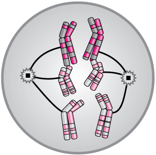
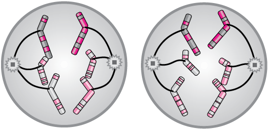
Anaphase I
In anaphase I one homologous pair is pulled toward each pole of the cell in preparation for dividing into two new diploid cells. These cells would be like any other normally functioning cell except that the crossing-over process has occurred.
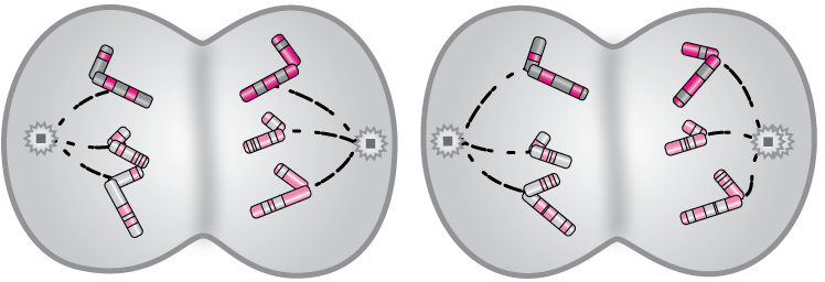
Telophase I
The first phase of meiosis ends with the two new cells pinching off and forming complete cell membranes. The spindle fibers dissolve,

yielding two new diploid cells with significant changes in the DNA sequence of the chromosomes, again represented here by the striping of the chromosomes.:

In meiosis I, a tetraploid cell (a diploid cell in which all chromosomes have been copied) is split into two diploid cells in which homologous chromosomes have swapped segments of DNA, resulting in genetic change.
Meiosis II: Forming four haploid gametes
Prophase II
Meiosis II begins where telophase I left off. We can call the end of telophase I prophase II. Chromosomes are still maximally condensed, and each cell is diploid, containing a homologous pair of each kind of chromosome.
Metaphase II
Metaphase II resembles metaphase of mitosis. Homologous pairs are lined up on the metaphase plate with spindle fibers attached to the centromeres, one to each homolog.
Anaphase II
During anaphase II, two diploid cells are converted into four haploid cells that will be gametes — sperm and egg cells of mammals. One homolog is segregated to each pole of the two diploid cells that came out of meiosis I
Telophase II
In telophase II, the cytoplasms of each cell divide, sharing contents, and the cell membranes pinch off to form four new cells, all haploid with new genetic diversity.
The result is four haploid gametes that will usually undergo further maturation in preparation for fusion with the gametes of a partner in sexual reproduction. That mixing (1 gamete + 1 gamete), plus the mixing of DNA from crossing over, yields the majority of the diversity we see from generation-to-generation in organisms.

In meiosis II two diploid cells are split into four haploid cells that will go on to form gametes. In sexual reproduction, gametes from each parent fuse to produce a new diploid cell that will be the progenitor of every other cell in the new organism.
Overview
Here is an overview of all of the steps of meiosis, plus some. Click/tap on the figure to download a .pdf copy of your own.


![]()
xaktly.com by Dr. Jeff Cruzan is licensed under a Creative Commons Attribution-NonCommercial-ShareAlike 3.0 Unported License. © 2016-2025, Jeff Cruzan. All text and images on this website not specifically attributed to another source were created by me and I reserve all rights as to their use. Any opinions expressed on this website are entirely mine, and do not necessarily reflect the views of any of my employers. Please feel free to send any questions or comments to jeff.cruzan@verizon.net.